Science with Sandra: New Publications

Welcome to Science with Sandra!
I hope you are enjoying Spring time! For the April edition of Science with Sandra, I would like to share a brief summary of two publications. The title of the first one is: “An arrayed CRISPR knockout screen identifies genetic regulators of GLUT1 expression”. This manuscript was published by Dr. Ethan Lippmann and his team in Vanderbilt University.
Before I start sharing information about the manuscript, I would like to give you a brief explanation of CRISPR and the technique used for this study. CRISPR Cas 9 is a laboratory tool used to edit pieces of DNA in a cell. It uses a specially designed RNA molecule to guide the enzyme Cas9 to a specific sequence in the DNA. The enzyme cuts the strand of targeted DNA and removes a small piece, leaving a gap. Then, the DNA that remains can be modified or a new piece of DNA can be added. In this link you can find a short video of this technique published by the Mayo Clinic.
The study from Dr. Lippmann and his team uses an arrayed CRISPR knockout screen in order to identify genetic regulators of GLUT1. This technique uses the CRISPR Cas9 system to reveal relationships between genotypes (or genetic information) and phenotypes (or the symptoms and what can be observed). Arrayed CRISPR screens allow identification of the effect of multiple gene knockouts (or gene inactivations or removals) on the phenotype of a cell.
The goal of this study was to have a better understanding of the regulation of the expression of the GLUT1 protein due to its importance in maintaining energy homeostasis and its implications in diseases such as Alzheimer’s, cancer and GLUT1 Deficiency. Dr. Lippmann and his team, explained how GLUT1 is key for the normal function of processes such as embryo development, glucose movement across the placenta, blood vessel formation in the central nervous system and activation of T cells, which are important players in the immune response.
The importance of understanding the regulation of GLUT1 is key in order to developing treatments not only for GLUT1 Deficiency, but also for other conditions such as Alzheimer’s and cancer.
For the current study, they used a cancer cell line that has a high expression of the SLC2A1 gene that encodes for the GLUT1 protein as a model to study GLUT1 regulation. In addition, they used an automated high content immunostaining approach to quantify GLUT1 expression. The team screened 8,000 genes and tested the effect on the removal of each of those genes in the expression of GLUT1. The results of the screen revealed 300 genes that when removed, led to the downregulation or reduced expression of the GLUT1 protein. Among the genes where removal led to the decreased expression are genes involved in signaling pathways activated by G protein-coupled receptors (GPCRs), which are integral membrane proteins that recognize a wide variety of signals including hormones and neurotransmitters.
Many of the GPCRs identified in their screen are predicted to be expressed on brain endothelial cells. Brain endothelial cells are part of the Blood Brain Barrier and the first cell type to come in close contact with blood and the molecules coming into the brain.
Interestingly, a deeper analysis showed that many of the genes identified in the study can be activated by small molecules and metabolites including catecholamines, acetylcholine, dopamine, adenosine, prostaglandins, sphingosines, and fatty acids. The authors discuss how the identification of catecholamine and dopamine receptors implies that emotional state can regulate GLUT1 expression, because catecholamines and dopamine are produced in response to stress.
The results of this study suggest that diverse signaling pathways can regulate GLUT1 expression. In addition this study opens up the possibility of exploring different mechanisms to increase or decrease the expression of GLUT1 for different conditions including GLUT1 Deficiency Syndrome.
The second publication I would like to highlight is: “Human In Vitro Models of Neuroenergetics and Neurometabolic Disturbances: Current Advances and Clinical Perspectives”. This manuscript was published by the group pf Dr. Anna Herland from the KTH, Royal Institute of Technology in Sweden.
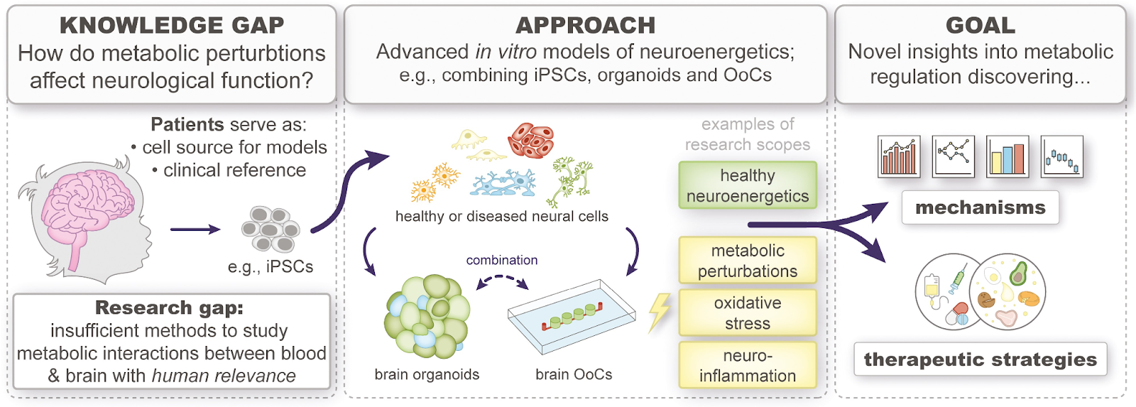
The goal of this study was to explore the key steps and considerations involved in developing an advanced human in vitro model for studying neuroenergetics or the central nervous system’s energy metabolism. The authors provided in the manuscript a graphical abstract of their manuscript which is really useful to understand their study (please see below).
Rogal J et al, 2024
The authors discuss how despite increased evidence of the important role of energy metabolism in brain function, there is a limited understanding of the mechanism of brain energy metabolism in humans due to the lack of adequate models to perform these studies. The number of neurological conditions has raised in the past decades, mostly on neurodegenerative diseases in the aging population.
Dr. Herland and her team explained the importance of better understanding the brain and brain energy metabolism. They emphasize the important role glucose plays not only as a source for ATP production, but also as a precursor of other molecules produced in the brain such as neurotransmitters.
Because the brain does not have a high glucose storage capacity, it needs to rely on a constant glucose supply from the blood through glucose transporters including GLUT1. GLUT1 is highly expressed in brain endothelial cells, and it has been shown that its expression decreases with aging.
With age, glucose transporters such as GLUT3 and GLUT4, which express in neurons in the brain, decrease their expression as well. Dr. Herland and her team, shared information regarding Alzheimer’s disease (AD), where the decreased expression of GLUT1, GLUT3 and GLUT4 can lead to cell membrane destabilization, DNA and protein damage.
“GLUT1-DS is an excellent point of reference in neuroenergetics, allowing for unprecedented insights into the role of glucose, and especially its transport.”
Dr. Anna Herland
Regarding GLUT1 Deficiency, the authors stated “GLUT1-DS is an excellent point of reference in neuroenergetics, allowing for unprecedented insights into the role of glucose, and especially its transport”. The statement adds to the thought shared by researchers in our community, that GLUT1 Deficiency is a prototype disease to study not only this condition but other conditions in which brain glucose metabolism is affected.
The figure below, taken from this publication, explains how the limited treatments available for neurological disorders put a burden in the healthcare system. The figure also depicts how neuroenergetics are disrupted in diseases such as AD, traumatic brain injury or GLUT1 Deficiency.


Rogal J et al, 2024
The authors of this publication, describe how research models to study the brain in health and disease don’t really capture the brain’s complexity. Some of those models include animal models and 2D human cell cultures. An alternative approach is the use of microphysiological systems (MPSs) that include organoids, which are miniaturized and simplified versions of organs that are self-assembled 3D tissue constructs and derived from stem or progenitor cells.
Other example includes the organ on a chip (OoC) model. In this model, living cells are cultured in micrometer-sized chambers that generate functional units of specific tissues. For this model, due to the precision in size and dynamic liquid exchange, highly controllable physicochemical and spatiotemporal parameters can be induced. Dr. Herland and her team illustrate the importance of human induced pluripotent stem cells (iPSCs) in both models to achieve their full potential. According to the authors, use of patient derived iPSCs will likely play an integral part of future precision medicine.


Rogal J et al, 2024
Finally, the authors describe different aspects that need to be taken into consideration in order to develop the best models to study brain metabolism in health and disease. Some of those aspects include the cellular complexity of the cells implicated in the topic of interest. In the case of GLUT1 Deficiency, it is known that endothelial cells, astrocytes and neurons are directly and indirectly impacted by the low expression of GLUT1; therefore, utilizing a model that involves each of those cells to study the impact of low GLUT1 expression will help to have a better understanding of the disease and to develop better treatments for this condition.
Another aspect to consider is the cell source and the topic being studied. The authors point out that when using patient derived iPSCs it is important to consider the age of the patient to see if the cell maturity state reflects the characteristics the cell has, especially when studying neuroenergetics in aging. For example, the use iPSCs cells derived from older adults to study AD rather than using cells derived from children, could be more representative model of what is actually happening.
Dr. Herland and her team mentioned that other important aspects to consider when studying neuroenergetics include the neurometabolic pathways involved in the condition that’s being studied, the physiological microenvironment, such as oxygen levels and nutrients present, as well as the imaging methods or tools used to monitor the metabolic process in real-time.
This publication describes different aspects that are important to consider when studying neurometabolic disorders and serves as a guide to develop better and improved models to study these disorders, which will take into consideration the complexity of the brain and its metabolism in health and disease.
Thank you for visiting our blog! Please do not hesitate to contact me at [email protected] if you have any questions or comments.

