
A Closer Look at a Select Group of Missense Variants

Hello and welcome to Science with Sandra!
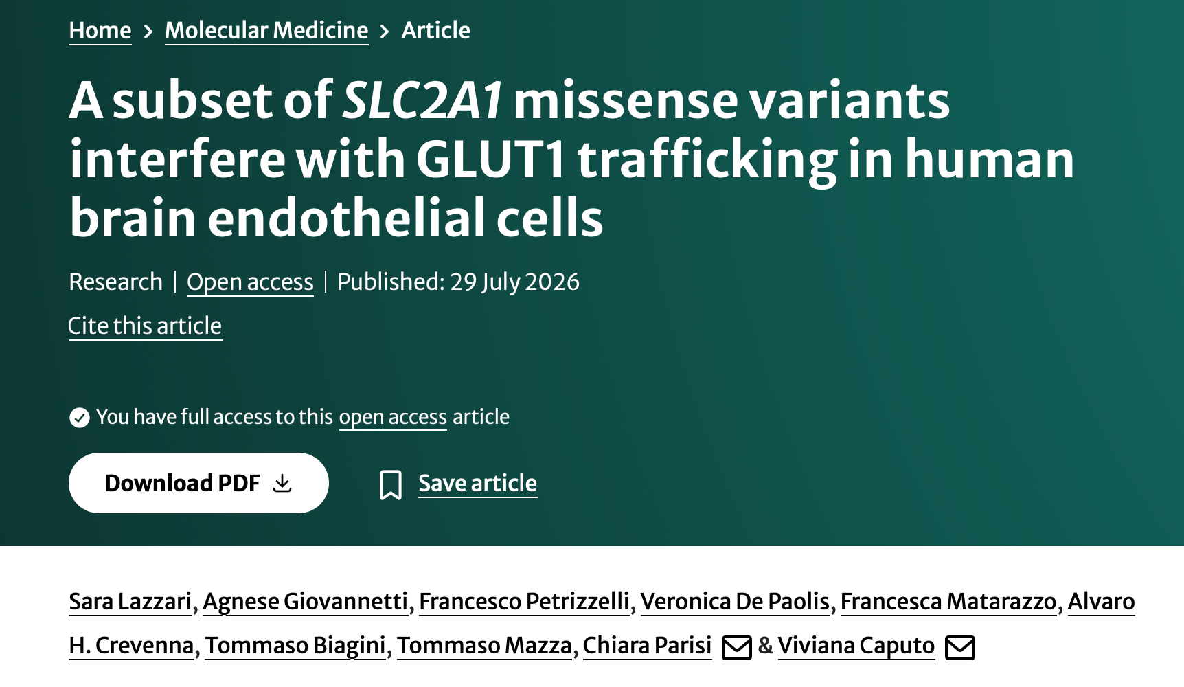
For this edition, I would like to share a recent publication by a group of researchers led by Dr. Viviana Caputo and Dr. Chiara Parisi in Italy. The title of the publication is “A subset of SLC2A1 missense variants interfere with GLUT1 trafficking in human brain endothelial cells.”
The goal of the study was to investigate a group of selected GLUT1 variants to assess their localization in the cell in response to changes in metabolic conditions.
What is important about this study?
Glucose transporter type 1 (GLUT1) is one of 14 glucose transporters in the body, but the only one present in the blood-brain barrier (BBB). This protein is present in erythrocytes (red blood cells), and in the brain it is present in endothelial cells and astrocytes. The brain depends almost entirely on glucose for fuel; therefore, GLUT1 is essential for normal brain function.
The gene that carries the instructions for making the GLUT1 protein is called SLC2A1. When this gene has certain changes, or variants, this can result in GLUT1 Deficiency. As many of you know, people living with GLUT1 Deficiency may experience a broad spectrum of symptoms with a broad spectrum of severity. The severity of symptoms varies widely from person to person, even among people who share the same gene variant.
The majority of SLC2A1 variants are missense, meaning there was a one-nucleotide change in the DNA that can cause a change in the amino acid that makes up the protein. In general, it is thought that these variants cause a milder phenotype, meaning the symptoms experienced by people with this type of variant are milder compared to people with other types of variants. It is also thought that these variants cause disease mainly by reducing how much glucose can be transported. This study asked whether some of these variants could also disrupt GLUT1 in other ways, specifically by preventing the protein from reaching the right location in the cell in the first place.
What did the researchers do?
The researchers studied eight different SLC2A1 variants chosen to represent different structural regions of the GLUT1 protein with different levels of clinical and functional effects. The pathogenic, or disease-causing, variants tested include p.N34S, p.R126L, p.R153C, p.R223P, p.R223W, p.Q283R, and p.P485L, along with one variant of unknown significance, p.I404V, which was included for comparison.
To study these variants, researchers attached a glowing green protein tag (GFP) to the healthy and variant versions of GLUT1 and introduced them into two types of human cells grown in the lab: brain endothelial cells and a widely used laboratory cell line called HeLa cells. To track each version of the GLUT1 protein inside the cell, they used specialized microscopy techniques. These allowed them to see where the protein was located, whether it reached the cell surface, where it normally functions to transport glucose, or whether it stayed inside the cell in the cytoplasm or another cell compartment, including lysosomes.
To identify the cellular compartments, the researchers used molecular markers, proteins specific to each compartment, which allowed them to determine the location of the GLUT1 protein. Researchers also tested how cells responded to different amounts of glucose and measured how quickly each variant of the GLUT1 protein was broken down and replaced over time.
What did the researchers find?
Four of the eight variants (p.R153C, p.R223P, p.R223W, and p.I404V) behaved essentially like the healthy version of the protein, in that they were able to reach the cell membrane. This does not mean that p.R153C, p.R223P, and p.R223W are harmless, it simply means that the GLUT1 protein produced with these changes has correct localization in the cell.
On the other hand, variants p.N34S, p.Q283R, p.R126L, and p.P485L did not reach the cell surface and were instead retained inside the cell. The first three of these accumulated inside lysosomes, the cellular compartments that break down and recycle cellular material, while the last variant accumulated in the Golgi apparatus and early endosomes. The Golgi apparatus is where proteins are prepared for shipping to their destinations in the cell, while early endosomes help sort proteins for recycling or for transport elsewhere in the cell.
Interestingly, when researchers looked at the variants localized in lysosomes, they found that the proteins were not being degraded efficiently, as it normally happen in lysosomes. Instead, these proteins remained there, as if stuck, without being cleared out.
The researchers also tested whether these localization issues changed depending on how much glucose was available to the cells. Normally, GLUT1 responds to glucose availability: when glucose is low, GLUT1 localizes to the cell membrane, while when glucose is high, it moves away from the cell membrane for degradation or storage. The results showed that the localization of variants p.N34S, p.Q283R, and p.P485L did not change even when glucose was low.
Finally, the research team compared their laboratory findings to information found in clinical databases, AI prediction models, and the existing literature. Clinical databases compile information from scientists and clinicians about genetic variants and whether they are disease-causing, not disease-causing, or of uncertain significance. There are also AI models that predict the pathogenicity of missense variants in humans. The researchers found discrepancies between their results and the predictions from these models. For example, some variants were described by AI models as low-risk or uncertain, but showed clear, significant abnormalities in the lab experiments, consistent with what the clinical databases indicated. These discrepancies highlight that computer-based predictions, while useful, cannot fully substitute for laboratory testing when it comes to understanding the functional effects of gene variants and their possible impact on patients.
What does this study mean for our community?
This study is relevant to our community because, first of all, it adds valuable information about SLC2A1 missense variants. Most importantly, it shows how heterogeneous missense variants can be, and that the effect of a variant may not simply be an inability to transport enough glucose, but could also derive from incorrect cellular localization.
The results of this study are important because they open new possibilities for drug development. Identifying the precise effects of a gene variant on a protein could help determine which existing or future medications might help correct protein localization, for example. The research team highlights the importance of follow-up studies using patient-derived models to determine whether these results are reproducible.
We thank Dr. Caputo and Dr. Parisi and their teams for all their hard work and for their interest in working and learning more about GLUT1 Deficiency.
Than you for visiting our blog and please do not hesitate to contact me at [email protected] if you have any questions.