
Disrupted Glial Metabolism and Myelin Vulnerability

Hello and welcome to Science with Sandra!
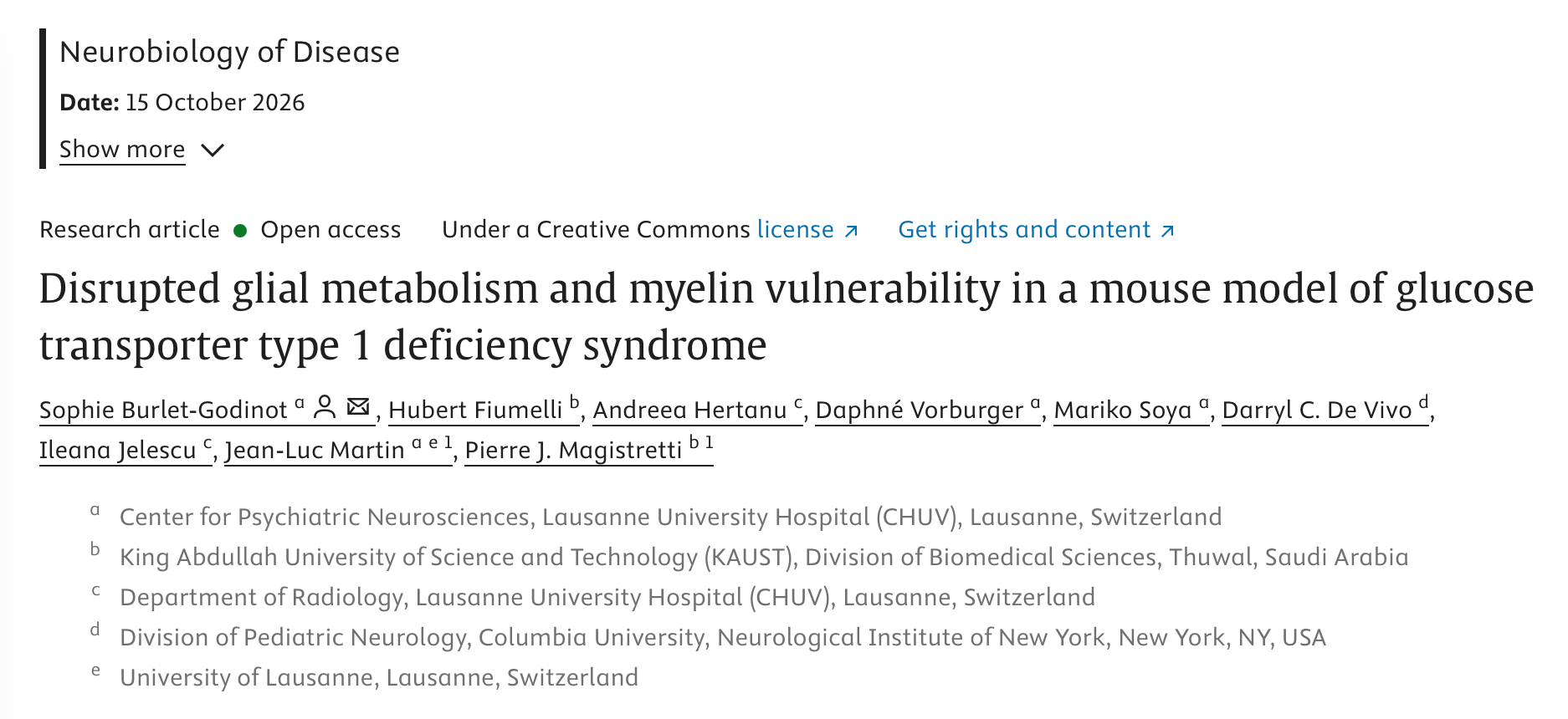
For this edition, I would like to highlight a recent publication by Dr. Pierre Magistretti, his team at King Abdullah University of Science and Technology, and their collaborators, including Dr. Darryl De Vivo. The publication is titled “Disrupted glial metabolism and myelin vulnerability in a mouse model of glucose transporter type 1 deficiency syndrome”.
GLUT1 Deficiency is caused by impaired glucose transport into the brain due to a reduction in the GLUT1 protein, which is the primary glucose transporter expressed in the endothelial cells of the blood-brain barrier, as well as in astrocytes and oligodendrocytes within the brain.
As a consequence, people with GLUT1 Deficiency can experience a broad spectrum of symptoms, including seizures, developmental delays, movement disorders, speech and language difficulties, and cognitive challenges. Not all patients experience all of these symptoms, and their severity varies from person to person.
The standard treatment for GLUT1 Deficiency is the ketogenic diet, which provides ketones as an alternative source of fuel for the brain. In the majority of cases, the diet works well, particularly for seizure control. However, approximately 10-15% of patients in the community do not respond adequately to the diet.
Currently, it is not fully understood why the ketogenic diet does not address all of the symptoms associated with GLUT1 Deficiency. The study by Dr. Magistretti and his team aimed to better understand the effects of chronic glucose shortage at the cellular level, specifically in astrocytes and oligodendrocytes.
Astrocytes are key cells in the brain that help support energy metabolism and communication between different types of brain cells. Oligodendrocytes are specialized cells that produce myelin, the fatty insulating layer surrounding neurons that allows neuronal signals to travel quickly and efficiently.
What did the researchers study?
Dr. Magistretti and his team used a mouse model of GLUT1 Deficiency. This model has only one functional copy of the SLC2A1 gene which encodes for the GLUT1 protein and reproduces several important features of the human disease, including reduced brain glucose uptake, low cerebrospinal fluid (CSF) glucose levels, motor abnormalities, and microcephaly.
The researchers performed a series of metabolic measurements and analyzed magnetic resonance imaging data, as well as molecular data, including RNA and lipid profiles. Their goal was to identify cellular pathways that are particularly vulnerable to chronic brain glucose deprivation and that could potentially inform new metabolic therapeutic strategies.
What did the researchers find?
As expected, the researchers found that glucose levels in the brains of Glut1-deficient mice were lower than those of healthy control mice. In addition, glycogen, an important energy reserve, was reduced, as was lactate, a key metabolite involved in brain energy metabolism.
Significant reductions were observed in multiple brain regions, but the changes were particularly pronounced in the striatum, a region of the brain that plays a central role in locomotor control. Within the striatum, the dorsolateral region is especially important for sensorimotor processing. These findings could potentially help explain why motor problems are an important challenge for many people with GLUT1 Deficiency.
When the researchers evaluated gene expression in the brain, they found that Glut1-deficient mice had reduced expression of genes involved in the formation and maintenance of myelin, particularly in the striatum. On the other hand, gene expression analyses also indicated that chronic glucose reduction induced changes in astrocytes that may help these cells adapt to an environment with reduced glucose availability.
Because of the critical role of astrocyte-oligodendrocyte connections in myelin formation and maintenance, the researchers also evaluated the expression of a specialized group of proteins called connexins, which allow direct communication between cells. Their results indicated that a subset of these proteins was significantly reduced in the striatum of Glut1-deficient mice, suggesting that communication between these cell types may be disrupted.
What about myelin and lipids?
Lipids are major components of myelin, accounting for approximately 70-80% of its dry weight. The researchers therefore performed a detailed analysis of lipid composition in the striatum.
They found that the relative abundance of specific lipid classes, including lipids associated with cellular membranes and myelin, was altered in the striatum of Glut1-deficient mice, despite the total lipid content remaining unchanged. This suggests that GLUT1 deficiency may cause a selective shift in the composition of lipids associated with myelin rather than simply reducing the overall amount of lipids in the brain.
Could lactate help?
Because of the reduction in lactate levels observed in the brains of Glut1-deficient mice, and because of the important role of lactate in brain energy metabolism and myelination, Dr. Magistretti and his team decided to test whether administering L-lactate to young mice could improve motor performance.
The results showed that L-lactate administration to young Glut1-deficient mice improved their motor performance without negatively affecting body weight, blood glucose, or blood lactate levels.
Dr. Magistretti and his team propose several possible explanations for these beneficial effects. First, lactate could contribute to the restoration of myelin integrity in addition to serving as an alternative energy source, by providing carbon that can be used for lipid synthesis. The researchers also suggest that lactate could potentially act as a signaling molecule involved in physiological processes important for neurodevelopment, as has been suggested by other studies. Another possibility is that lactate may help support the integrity of the brain’s blood vessels.
Why is this study important for our community?
It provides a potential biological explanation for motor symptoms experienced by people living with GLUT1 Deficiency.
Many families know that movement difficulties can persist, or even worsen, despite good adherence to the ketogenic diet. This study suggests one possible explanation: GLUT1 Deficiency may affect myelin composition and the function of brain regions involved in locomotor control, processes that may not be fully addressed by the ketogenic diet alone.
It puts myelin on the map as an important area for future research.
The role of myelin and the effects of reduced brain glucose metabolism on myelin have not received as much attention as other aspects of GLUT1 Deficiency. This study highlights the importance of investigating this area further and raises the possibility that lactate administration, particularly early in life, could eventually be explored as a therapeutic strategy.
It is important to remember that these findings are still at the preclinical stage.
The researchers emphasize that more work is needed to understand the mechanisms underlying the beneficial effects of lactate. Additional research will also be necessary to determine whether these findings can be translated into a potential therapeutic strategy for people with GLUT1 Deficiency and, ultimately, whether this approach could be evaluated in clinical trials.
Finally, this study corroborates findings from other researchers in our community who observed decreased brain glycogen levels in a different mouse model of GLUT1 Deficiency. Together, these findings strengthen our understanding that GLUT1 Deficiency is not simply a seizure disorder or an energy-deficit disorder. Rather, impaired glucose transport may disrupt the metabolism and function of multiple types of brain cells, contributing to the broad spectrum of symptoms experienced by people living with the disease.
This paper also expands our understanding of potential therapeutic strategies that could, with further research, benefit people living with GLUT1 Deficiency.
We thank Dr. Magistretti, his team, and their collaborators for their important work and for their continued interest in helping our community.
Thank you for visiting our blog and please do not hesitate to contact me at [email protected] if you have any questions.