Science with Sandra

Hello and welcome to Science with Sandra! This time I would like to share a brief summary of a recent publication by Professor Dr. Katrien De Bock and her team at the Laboratory of Exercise and Health at the Swiss Federal Institute of Technology in Zurich, Switzerland. Dr. De Bock. The title of her publication is “Endothelial metabolic control of insulin sensitivity through resident macrophages”.
The goal of this publication was to have a better understanding of the role of endothelial cells (ECs) in muscle glucose homeostasis.
In this article, Dr. De Bock and her team explain how skeletal muscle plays a significant role in maintaining the balance of whole-body glucose or homeostasis. The authors mention that during rest, around 25%-45% of all ingested glucose of a mixed meal is taken up by the muscle. Glucose and other nutrients are delivered to the muscle by endothelial cells lining up blood vessels carrying these nutrients. Endothelial cells not only transport insulin, but can also enhance glucose delivery to muscles through different mechanisms. Similar to the brain, skeletal muscle ECs express GLUT1, however in contrast to brain, in skeletal muscle glucose enters in between the endothelial cells, so the role of GLUT1 in muscle ECs is not known.
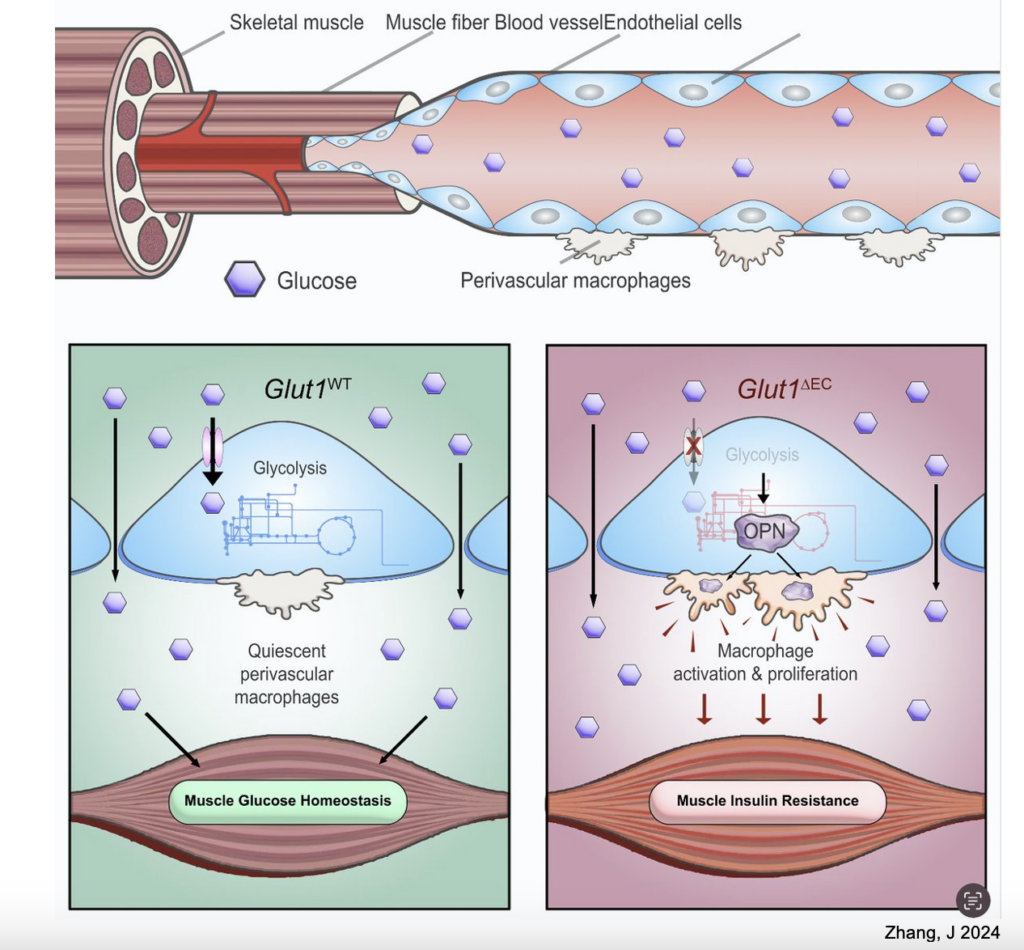
Dr. De Bock and her team used a mouse model where GLUT1 was absent in endothelial cells to determine if GLUT1 was important for the transport of glucose into muscle tissue. Their results confirmed that it is not required to have active transport through GLUT1 for glucose to move into skeletal muscle. However, the absence of this GLUT1 transporter in ECs induces muscle insulin resistance. Muscle insulin resistance can eventually lead to reduced muscle mass, muscle strength and function.
Why does loss of endothelial cell GLUT1 induce muscle insulin resistance? Well, the authors found that the loss of GLUT1 in endothelial cells leads to the activation and increase in the number of muscle macrophages residing in skeletal muscle. Those macrophages lie close the endothelial cells in the muscle. Interestingly, authors determined that GLUT1 expressed by endothelial cells controls muscle insulin sensitivity by controlling the accumulation of these macrophages through the secretion of osteopontin (OPN) from endothelial cells. Osteopontin is a protein that has many different functions including cell migration, adhesion, and in the immune response.
In addition to this finding, researchers found that loss of GLUT1 in ECs reduces glucose uptake into endothelial cells which rewires their glucose metabolism leading to the activation of Osteopontin. How this works still need further investigation. What is also interesting is that the intake of a high-fat diet affects GLUT1 levels in endothelial cells.
Finally, the authors mention how the loss of GLUT1 in brain ECs leads to decreased glucose transport across the blood-brain barrier. The authors found that in contrast to this, the loss of GLUT1 expression in muscle ECs does not impair glucose transport across the endothelial barrier in skeletal muscle, but controls glucose uptake into endothelial cells. The authors suggest that the transport of glucose across the endothelial cells in skeletal muscle occurs via other routes besides the GLUT1 transporter, with the purpose of maintaining the necessary glucose metabolic balance or homeostasis in this tissue.
Below you can see a figure from her publication that summarizes their findings.
We thank Dr. De Bock and her team for her studies to have a better understanding of the role of the GLUT1 transporter and for taking the time to review this summary.